Single-Cell Seeding: From Basics to Advanced Methods
The single-cell seeding process differs across laboratories and institutions, and there are many ways to perform single-cell seeding depending on the resources and equipment available1. While these methods can produce clonally distinct populations growing in a single well, they come with varying degrees of efficiency, risk, and consistency. Single-cell seeding is inherently challenging as it requires the addition of a single cell to a single well. Thus, the choice of method can significantly impact the success of a research project and the speed at which a potential therapeutic traverses the drug discovery pathway. Here, we discuss the different methods of single-cell seeding and monitoring, highlighting the relevant advantages and disadvantages and looking to the future of the single-cell seeding process.
Seeding
Accurate seeding of single cells into a single well underpins the success of many modern therapeutics. Indeed, cell-based therapies and interventions such as antibodies that require a cell to be produced begin with establishing a clonal cell population (Fig. 1)2. Modern regulatory standards require stringent documentation and proof of clonality3, which is harder to achieve with some seeding methods than with others. Common methods used for single-cell seeding include:

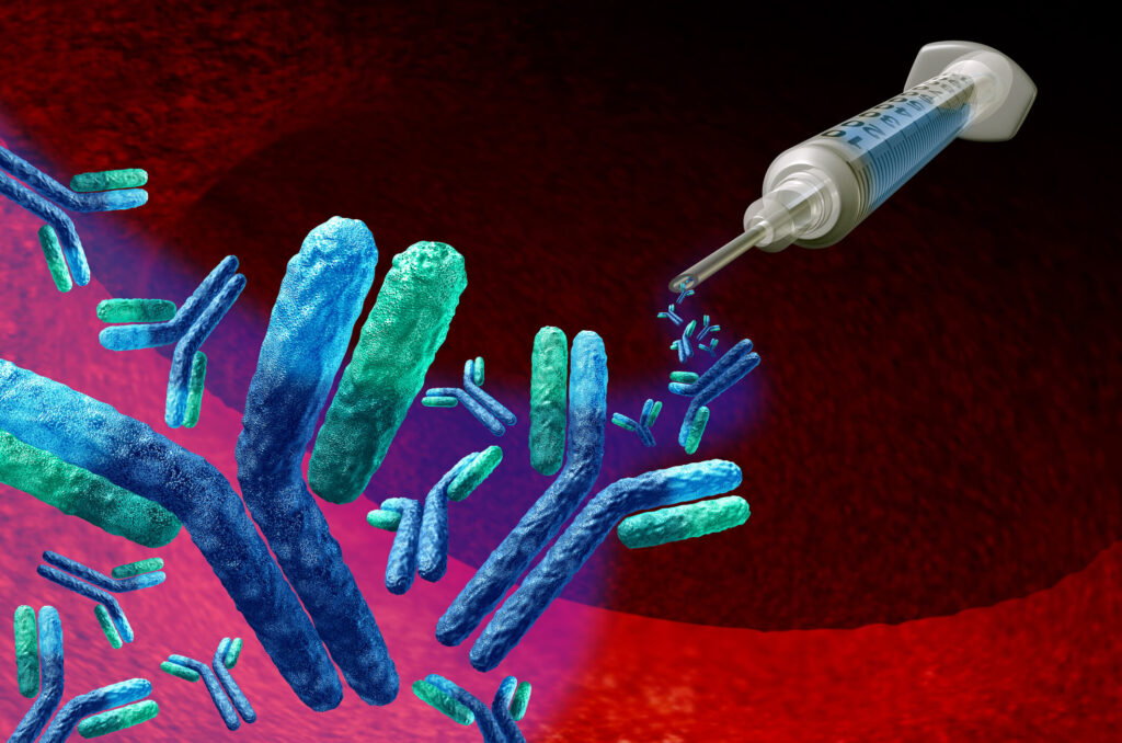
Figure 1. Monoclonal antibodies are used to treat a wide range of diseases including cancer and autoimmune diseases, and each antibody requires a verified clonal cell line for production.
Limiting Dilution
This method for single-cell seeding requires diluting cells to a sufficiently low concentration that when the suspension is added to a well, there is a higher probability of single-cell dispensing4. As such, this method does not guarantee a single cell per well but will also produce empty wells and wells with more than one cell. Limiting dilution does not require specialized equipment. However, it lacks the efficiency and reproducibility of more modern techniques.
Fluorescence-Activated Cell Sorting (FACS)
FACS is a type of flow cytometry that can sort heterogeneous mixtures of cells into different populations based on their size, granularity, and fluorescence characteristics. FACS machines can also be calibrated for single-cell dispensing into multiwell formats. Some cell types, like induced pluripotent stem cells, are sensitive to the pressure changes and shear forces experienced during flow cytometry. This limits the utility of FACS for some single-cell dispensing applications.
Microfluidics
Microfluidics technology facilitates the manipulation of small volumes of fluids through micrometer-scale channels, enabling close control over cellular environments, high-throughput analysis, and precision in cell dispensing5. Although microfluidics systems are more expensive than conventional equipment, they are far more efficient and robust in terms of reproducibility. These systems are now commonly found in automated liquid handling machinery, which offer significant advantages over conventional manual methods.
Automated vs Manual
Automation offers significant advantages over manual methods in the single-cell seeding process. This is especially evident when automation is coupled with microfluidic systems.
Efficiency: Automation is faster than manual methods and can help to streamline research projects. Automated dispensing runs can be performed overnight without the need for researcher supervision, which can increase research output severalfold.
Lower Risk: Manual methods of single-cell seeding increase the chances of human error, which comes with significant risks. Contamination can occur from two clones entering the same well and from microbial sources like mycoplasma. Automation eliminates the chances of contamination.
Researcher-friendly: With automation, researchers don’t have to spend time manually pipetting wells, increasing their risk of repetitive strain injury. Furthermore, automation frees up researchers' time for other work, such as interpreting results or designing the following experiment.
Accuracy: Automation is more accurate than manual pipetting. Over long periods, the chances for human error increase while automation remains consistent. Once an effective protocol has been established, an automated system can repeat the exact protocol precisely at any time. This ensures consistency both within laboratories and across different institutions.
Cost: Automated systems require a substantial upfront investment compared to manual pipetting. However, they offer significant savings over time. Speed is critical in the competitive drug discovery market, and automated processes can give researchers an edge over the competition.
Scalability: With microfluidics and automation, single-cell seeding can be scaled down to save money on reagents. Automated processes can also seed many clones in a fraction of the time compared to manual methods, allowing researchers to scale up their research as necessary.
Verifying Clonality
Regulatory bodies require robust evidence that a cell line used as a therapy or to produce a therapy was generated from a single clonal population3. Thus, verifying clonality after single-cell dispensing is essential for ensuring success in drug discovery. Manual inspection of wells using a light microscope is time-consuming and error-prone, making it challenging to prove clonality to regulatory bodies. Ultimately, researchers using this method may need to pay for expensive next-generation sequencing to confirm their population is monoclonal.
The UP.SIGHT all-in-one single-cell dispenserfrom CYTENA uses a dual imaging system to ensure single-cell dispensing and makes it easy to prove clonality to regulatory bodies (Fig. 2). The UP.SIGHT takes an image during cell dispensing and when it is successfully seeded into the well. This advanced method saves hours of researcher time and eliminates the chance of false positives with a > 99.999% probability of monoclonality.

Figure 2. The UP.SIGHT’s imagining system enables a >97% single-cell dispensing efficiency, and its small footprint means it fits easily inside most biosafety cabinets.
Monitoring
After successful single-cell seeding, researchers must monitor cells carefully to select promising clones. Clonality alone does not mean the cell will proliferate quickly or produce a high target protein yield. There are different methods for monitoring cells, including manual inspection using a light microscope. However, advanced imaging methods using the UP.SIGHT from CYTENA automatically tracks cell proliferation over time and provides researchers with images and crucial insights into cell proliferation and well confluency (Fig. 3).

Figure 3. The UP.SIGHT provides precise cell counts via integration with the advanced C.STUDIO software from CYTENA.
In many cases, cell proliferation is not sufficient for establishing good hits. Determining the titer of target proteins produced by cells is often essential. The F.QUANT from CYTENA offers a quick and robust way to test the concentration of antibodies in a biological sample, making it ideal for selecting the best hits to pursue.
Conclusion and Future Technologies
Single-cell seeding is pivotal in cell-based therapies and drug discovery, where precision and reproducibility are paramount. While traditional methods like limiting dilution remain cost-effective to an extent, advanced techniques such as microfluidics and automation provide superior accuracy and efficiency. Improvements in automation and the integration of image-based analysis and hit selection will continue to streamline the single-cell seeding process. The rapid development of single-cell multi-omics and real-time monitoring technologies will provide deeper insights into cellular behaviors and interactions, enhancing our ability to select therapeutic clones. Cumulatively, these advancements will streamline the development of cell- and biologics-based therapies to treat various diseases.
Ready to take your place at the cutting edge of cell-based therapies? Get in touch with one of our experts today to book a demo for the UP.SIGHT all-in-one single-cell dispenser and imaging system.
References
- Gross A, Schoendube J, Zimmermann S, Steeb M, Zengerle R, Koltay P. Technologies for Single-Cell Isolation. Int J Mol Sci. 2015;16(8):16897-16919. doi:10.3390/ijms160816897
- Lu RM, Hwang YC, Liu IJ, et al. Development of therapeutic antibodies for the treatment of diseases. J Biomed Sci. 2020;27(1):1. doi:10.1186/s12929-019-0592-z
- World Health Organization. WHO Expert Committee on Biological Standardization. World Health Organ Tech Rep Ser. 2013;(979):1-366, back cover.
- Staszewski R. Cloning by limiting dilution: an improved estimate that an interesting culture is monoclonal. Yale J Biol Med. 1984;57(6):865-868.
- Duncombe TA, Tentori AM, Herr AE. Microfluidics: reframing biological enquiry. Nat Rev Mol Cell Biol. 2015;16(9):554-567. doi:10.1038/nrm4041


